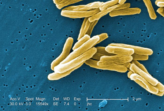
Under a high magnification of 15549x, this colorized scanning electron micrograph (SEM) depicted some of the ultrastructural details seen in the cell wall configuration of a number of Gram-positive Mycobacterium tuberculosis bacteria. 31 Oct 2014|Natalie Sambhi SHARE Share to Facebook Share to Twitter Share to LinkedIn Share to Email Print This Post With ImagesWithout Images Share SHARE Share to Facebook Share to Twitter Share to LinkedIn Share to Email Print This Post With ImagesWithout Images